X-ray optics: Transmission diffractive patterns of large microchannel plates
DOI:
https://doi.org/10.1109/ICATT.2017.7972589Keywords:
X-ray, X-ray optics, capillary, channeling phenomena, X-ray emission and fluorescenceAbstract
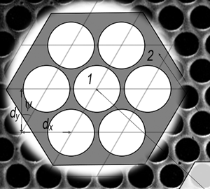
In this contribution we compare experimental and theoretical diffractive data of Micro Channel Plates (MCPs) in transmission mode. We evaluate the transmission efficiency of different optical devices at different energies of the primary X-ray radiation in the normal incidence geometry. Data were collected performing angular scans of both the MCP device and of the detector in the range of a few degrees. We analyzed MCPs of 33 mm and 20 mm diameter and ~300 μm thickness, having circular micro-channels of 3.4 μm directed normal to the MCP surface. Quite symmetric patterns of increasing complexity from high to low photon energy have been collected. Their shape and intensity are in reasonable agreement with preliminary simulations.References
K. Tsuji, J. Injuk, R. Van Grieken (eds.), X-Ray Spectrometry: Recent Technological Advances. Wiley, 2004.
Qi Zhang, Kun Zhao, Jie Li, Michael Chini, Yan Cheng, Yi Wu, Eric Cunningham, and Zenghu Chang, “Suppression of driving laser in high harmonic generation with a microchannel plate,” Opt. Lett., vol. 39, no. 12, p. 3670-3673, 2014. DOI: http://doi.org/10.1364/OL.39.003670.
M. A. Kumakhov, F. F. Komarov, “Multiple reflection from surface X-ray optics,” Phys. Rep., vol. 191, no. 5, p. 289-350, 1990. DOI: http://doi.org/10.1016/0370-1573(90)90135-O.
D. H. Bilderback, S. A. Hoffman, D. J. Thiel, “Nanometer spatial resolution achieved in hard x-ray imaging and Laue diffraction experiments,” Science, vol. 263, no. 5144, p. 201-203, 1994. DOI: http://doi.org/10.1126/science.8284671.
S. B. Dabagov, M. A. Kumakhov, S. V. Nikitina, V. A. Murashova, R. V. Fedorchuk and M. N. Yakimenko, “Observation of Interference Effects at the Focus of an X-ray Lens,” J. Synchrotron Rad., vol. 2, p. 132-135, 1995. DOI: http://doi.org/10.1107/S0909049595003864.
S. B. Dabagov, “Channeling of neutral particles in micro- and nanocapillaries,” Phys.-Usp., vol. 46, no. 10, p. 1053-1075, 2003. DOI: http://doi.org/10.1070/PU2003v046n10ABEH001639.
http://www.xos.com/focusing-optics.
H. N. Chapman, K. A. Nugent, S. W. Wilkins, and T. J. Davis, “Focusing and collimation of X-rays using microchannel plates: An experimental investigations,” J. X-ray Sci. Technol. Vol. 2, no. 2, p. 117-126, 1990. DOI: http://doi.org/10.3233/XST-1990-2203.
P. Kaaret, P. Geissbühler, A. Chen, and E. Glavinas, “X-ray focusing using microchannel plates,” Appl. Opt., vol. 31, no. 34, p. 7339-7343, 1992. DOI: http://doi.org/10.1364/AO.31.007339.
H. N. Chapman, K. A. Nugent, and S. W. Wilkins, “X-ray focusing using cylindrical-channel capillary arrays. 1. Theory,” Appl. Opt., vol. 32, no. 31, p. 6316-6332, 1993. DOI: http://doi.org/10.1364/AO.32.006316.
A. N. Brunton, MCP Optics. Thesis submitted to the University Leicester for Degree of Doctor Philosophy, Dec. 1994.
J. P. Nussey, Terrestrial and Space-based Applications of Microchannel Plate X-ray Optics, Thesis submitted to the University Leicester for Degree of Doctor Philosophy, July. 2005.